-
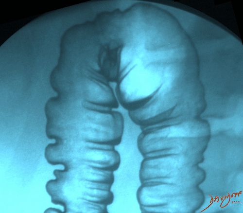
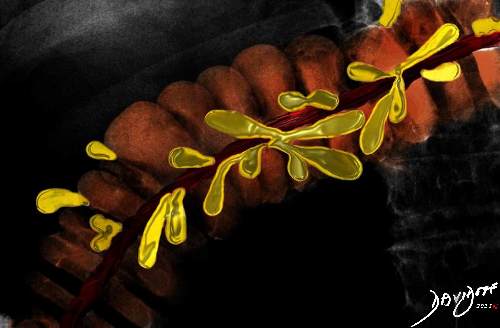
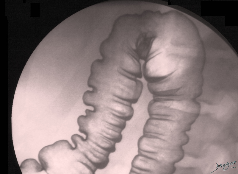
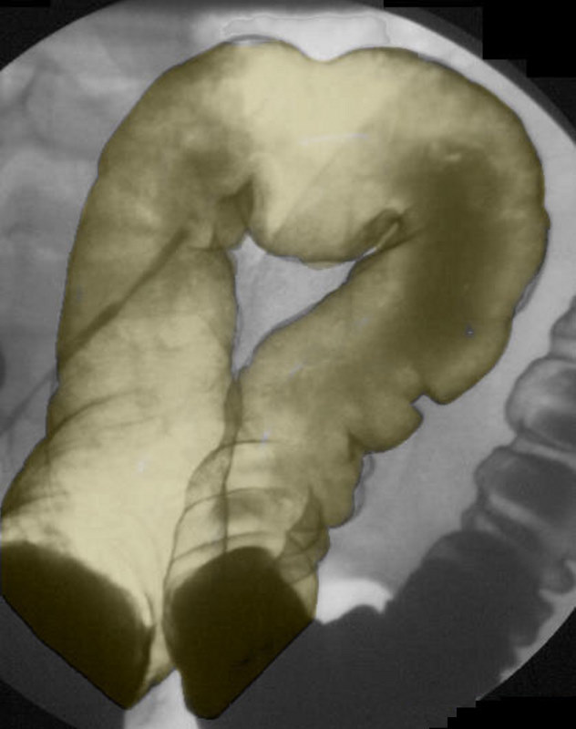
Descending Colon
| Parts of the Colon – Introduction
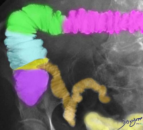
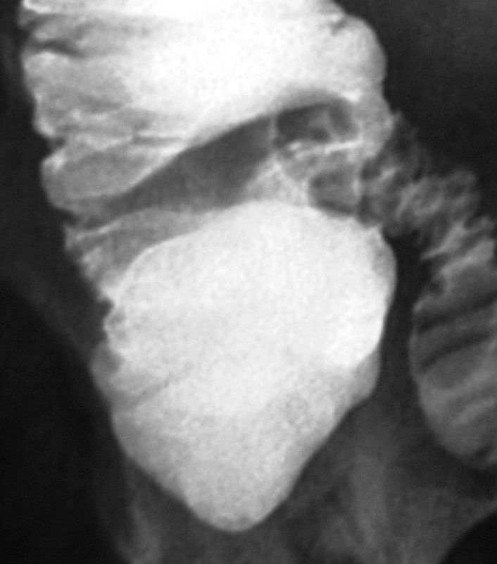
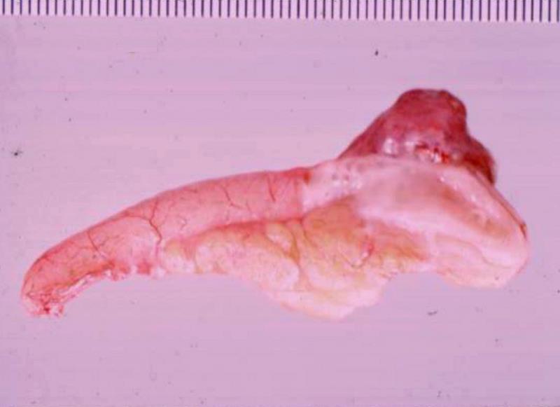
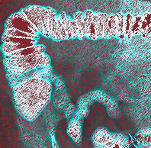
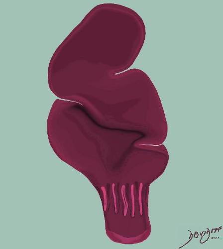
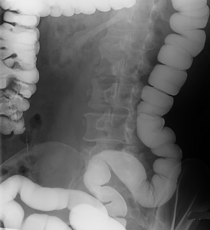
The colon is made up proximally of the cecum which is the widest potion which links the large bowel to the small bowel at the ileocecal junction. It contains the appendix which is a tubular structure about 3-5 mms in diameter and between 3 and 10cms long. Thereafter comes the ascending colon which is situated in the retroperitoneal position, followed by the transverse colon. The transverse colon hangs like a washing line within the peritoneal cavity between the two flexures – hepatic flexure on the right and the splenic flexure on the left. The descending colon is also in a retroperitoneal position while the sigmoid subtended on the sigmoid mesocolon becomes intraperitoneal. The rectum is bound down by the peritoneum as it exits at the anus.
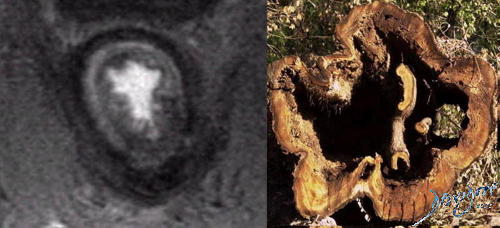
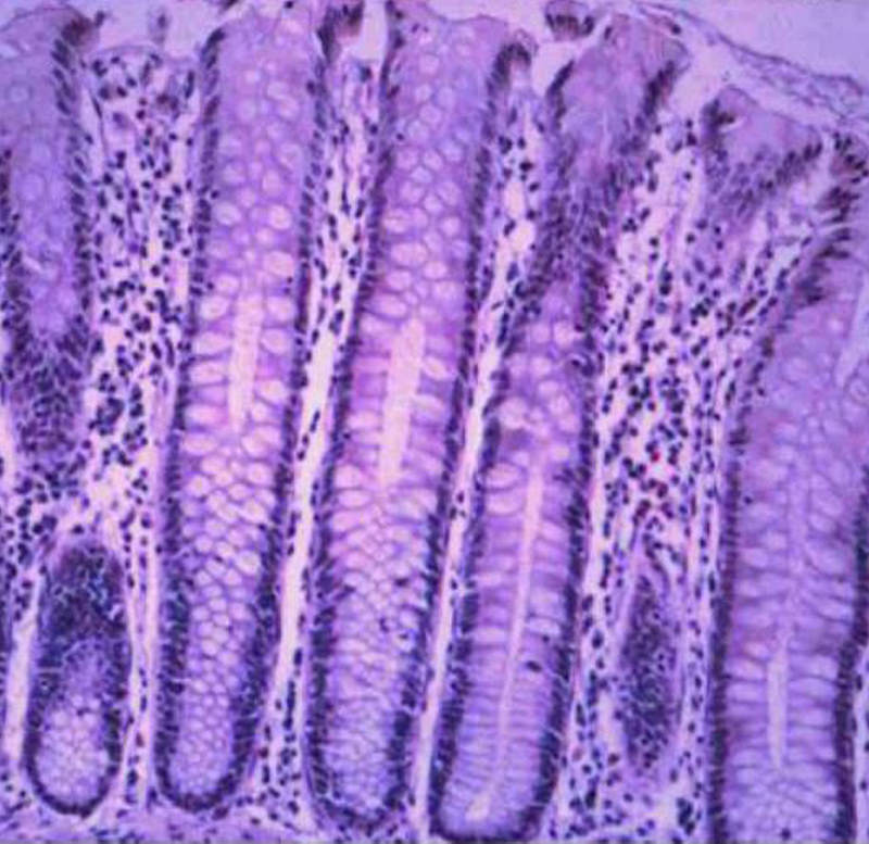
Each component of the colon has an inner endothelium, a submucosa, a middle muscular layer called the muscularis and an outer layer called a serosa or adventitia. This structural makeup was discussed in detail in the histology section. Minor but characteristic components include the taenia coli, appendices epiploica.
Support structures of the Colon
The mesocolon supports the intraperitoneal components while the retroperitoneal components are bound down by the peritoneum and are attached by loose areolar tissue to the retroperitoneal tissues. Ligaments include the transverse mesocolon, greater omentum, sigmoid mesocolon, mesoappendix, phrenicocolic ligament. The transverse mesocolon supports the transverse colon, and extends from the pancreas to posterosuperior asspect of the transverse colon. The gastrocolic ligament extends from the stomach to the superior aspect of the transverse colon. The sigmoid mesocolon supports the sigmoid colon and the mesoappendix supports the appendix.
The phrenicocolic ligament supports the splenic flexure by attaching to the diaphragm. It is a horizontal fold of peritoneum. The ascending and descending colon have no mesentery but are attached to the retroperitoneum by loose areolar tissue to the muscles such as the diaphragm, psoas, quadratus lumborum and the iliopsoas. |

