|
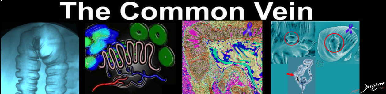
The appendix is a long narrow, blind ended tube that is also called the vermiform appendix because it has a worm like appearance (vermiform means worm-like in Latin). Its overall diameter is between 3-6mms when empty and its length ranges from 2-10cms, but can be up to 20cms. In the human appendix, the submucosa does contain lymphoid tissue and therefore the appendix is assumed to have a minor function within the immune system, though it has no role in digestion. It has a very narrow lumen and if this lumen becomes blocked from fecal material or due to lymphoid hyperplasia in the wall, secondary obstruction, dilatation, ischemia and infection results in the clinical entity of appendicitis. Weakening of the wall can be complicated by rupture with consequences of abscess formation or generalized peritonitis. The position of the appendix is variable and it can sometimes be very difficult to identify by CT, particularly when the normal appendix is not surrounded by fat and is instead surrounded by other loops of small and large bowel, or when the cecum itself is in an abnormal position. When the appendix is not obvious on initial perusal it is worthwhile using the more easily identified ileocecal valve with its fat laden lips as a guide, and then looking posteromedially and inferiorly for the appendix which usually lies within 2.5cms. of the ileocecal valve. If the tenia coli were easy to see on CTscan, finding the appendix in any situation would be a cinch since these longitudinal muscles all arise from around the appendix . These muscles are difficult to see on the CTscan.
Once the origin is identified, the body and tip of the appendix may end up posteriorly in a retrocecal position and can be superiorly directed in this position to the right, posterior or to the left of the cecum. On the other hand it may course inferiorly where it can also end up inferolaterally directly inferiorly or inferomedially. Occasionally it also found anteriorly. The appendix is retrocecal or retrocolic in about 65% of patients, pelvic in 30%, and inferior in about 4% and rarely anterior. (1%). The appendix is supplied by the appendicular artery, which is a branch of the anterior cecal artery which arises from the ileocolic artery. The appendicular artery can be found posterior to the terminal ileum where it finds its way into the mesoappendix before entering the substance of the appendix.
Each wall of the appendix can measure up to 3mms so that the total thickness (diameter) of the empty appendix should not be more than 6mms. If the wall cannot be separated from the contents then a diameter of up to 10mms is acceptable, (Benjaminov), though in practice even when the appendix does not reach these proportions the sense of the fluid filled distended appendix based on its shape and findings such as periappendiceal changes, and mucosal enhancement help clinch the diagnosis.
Applied Anatomy
CT scanning is highly sensitive and specific for the entity of appendicitis. Periappendiceal stranding is the most specific sign occurring in 100% of patients with the disease, while thickening of greater than 6mms is also sensitive (93%) and specific (100%) (Rao). The finding of appendicoliths on the other hand does not necessarily imply acute appendicitis, but is often associated with the acute condition. A stone stuck in the distal right ureter can sometimes masquerade as an appendicolith. ( Lane )
In humans the appendix is a vestigial structure, while in obligate herbivores the appendix is large, allowing it to participate in the fermentation process of cellulose. |