| Venous Drainage
The Common Vein Coyright 2007 IntroductionThe final common pathway of colonic venous drainage is dominantly via the portal system but also via the systemic venous system using the internal iliac veins to gain access to the IVC. The veins in general follow the course of the arteries and are similarly named. Thus there is a superior mesenteric vein (SMV), inferior mesenteric vein (IMV) and rectal or hemorrhoidal veins. The SMV usually drains with the splenic vein into the portal vein and in fact their confluence gives rise to the portal vein. The IMV usually drains into splenic vein. The middle rectal vein and inferior rectal vein drain into the internal iliac veins and then into the IVC as stated above.
Figure 1
There are two venous plexuses in the anorectal region. The superior hemorrhoidal venous plexus lies above the pectinate line and is covered with mucosa. The inferior hemorrhoidal venous plexus lies below the pectinate line, and is covered by anoderm and perianal skin. The superior plexus drains into the superior rectal vein while the inferior drains into the middle and inferior rectal veins. Applied AnatomyThere are two types of hemorrhoids – internal and external. The internal hemorrhoids are caused by varicosity and distension of the internal plexus while external hemorrhoids are caused by similar changes in the external plexus. Both are caused by increased intraabdominal pressure that may be caused by chronic constipation, obesity, pregnancy. They can also be caused by venous hypertension as might be related to portal hypertension.
|
 Mesenteric veins
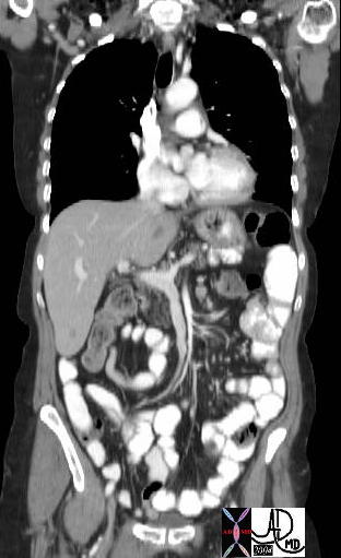
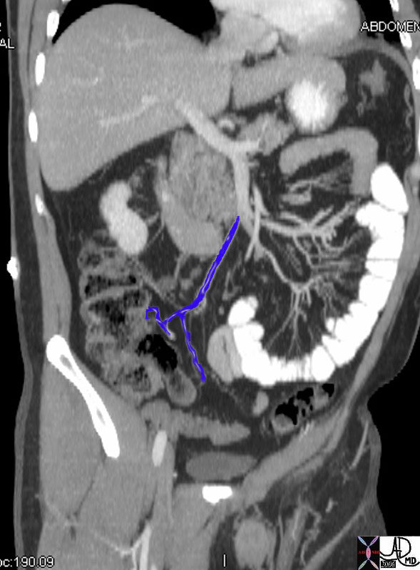 Figure 2
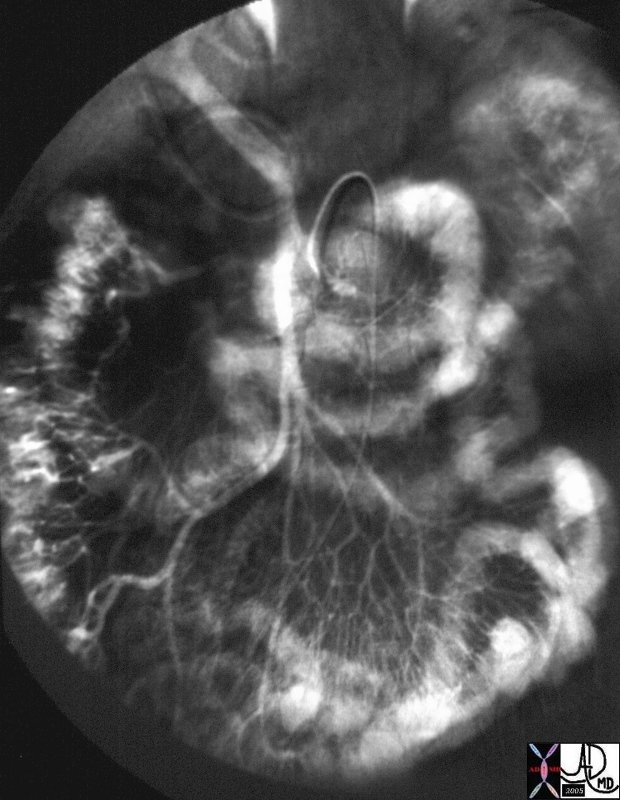 SMV and portal vein
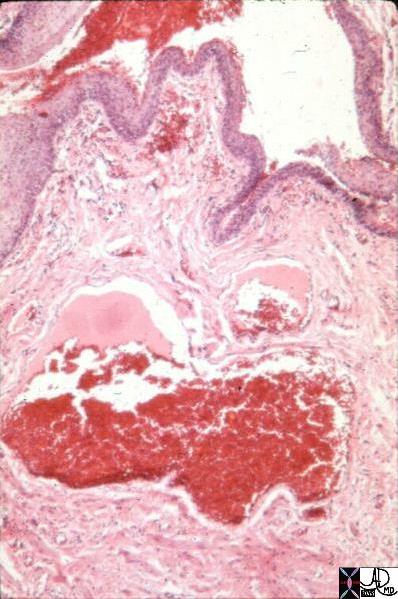 Hemorrhoids