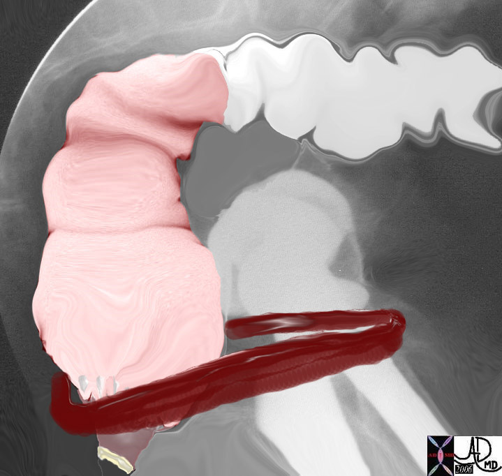
Anus
The anus is the last part of the colon. It consists of the anal canal which ends in the anal opening on the outside of the body. Anus means ring in Latin and it describes the shape of the organ. There are two sphincters that extend across the anal length which is about 3cms.. The internal sphincter is the involuntary smooth muscle that is found in close association with the pectinate line and is situated more proximally. The external sphincter is a voluntary skeletal muscle and it is found along the anal canal and in close association with the external opening and therefore situated more distally. These muscles in the basal state are both contracted until the act of defecation. Thus the anal canal is closed by muscle contraction in its “resting” state. The anorectal angle is the angle of the axis of the rectum with the axis of the anus which is produced by yet another group of muscles that affect anorectal function. At rest the normal angle is between 90 and 110 degrees. This angle is kept in position by the puborectalis muscle which acts as a sling around the anorectal junction. The puborectalis is attached anteriorly to the pubic bones and posteriorly it forms its sling around the anorectal junction. It is contracted under basal conditions so the anorectal junction is pulled forward creating the described 90-110 degree flexure. This angulation helps maintain continence. During defecation the puborectalis relaxes, the angle is reduced and the passage is straightened and opened. This function occurs in concert with the relaxation of both the internal and external sphincters causing the canal to open gaining a passage for the feces to enter the free world…. but that is yet another story.
The pelvic diaphragm is a hammock of skeletal muscle through which the pelvic viscera including the rectum pass. It is made of three muscles including the pubococcygeus, the ileococcygeus and the coccygeus muscle. The pubococcygeus is the anterior member, the ileococcygeus is in the middle and the coccygeus is posterior. The puborectalis described above is part of inner portion of the pubococcygeus. The pubococcygeus and the ileococcygeus together form the levator ani muscles. When feces enter the rectum the urge to defecate is initiated. The rectal distension results in relaxation of the involuntary smooth muscle of the internal sphincter. If the time is opportune the squatting or sitting position helps increase the intraabdominal pressure which pushes the pelvic diaphragm downward causing straightening of the anorectal angle. The voluntary signal to go forward and onward results in relaxation of the external sphincter and puborectalis and the anorectal angle is further straightened. Valsalva maneuver causes increased abdominal pressure which serves to flatten the levator ani and pelvic diaphragm even more, further providing a straightened anorectal angle and a straight shot. Manually pushing on the abdomen also helps increase the abdominal pressure aiding in the pushing down of the pelvic diaphragm. Signals about the activities in the smooth and skeletal muscles get transmitted to the autonomic nervous system and somatic nervous system resulting in mass peristalsis. The rectosigmoid is motivated to express itself while the transverse and descending colon are encouraged to do the same. The final sluice gate of the external sphincter needs to relax so that the event can happen. |
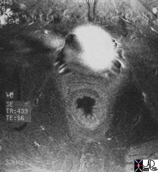 Anorectal region