|
The rectum is a remarkable organ with its ability to detect the nature of its contents enabled by a rich supply of nerves and blood vessels. It can for example distinguish between solid stool, liquid stool, and air. It is far more complex in form, sensitivity, and muscular function than any other part of the colon. The rectum is formed at about the 3rd sacral segment. As the sigmoid loses its freedom and dives back into the retroperitoneum to form the rectum, the three taenia coli expand to form a complete circumferential layer of longitudinal muscle rather the three distinct bands. The rectum ends at the anorectal junction where it turns at an angle of about ninety degrees posteriorly at the perineal flexure into the anus. Since there is no taenia coli in the rectum there are no haustra. Its walls are therefore relatively smooth but for three mucosal folds or valves that cross about one third of the diameter of the rectum.– two to the left side and one to its right. They are named according to their position as superior, middle and inferior rectal folds. The crescentic mucosal folds are also called Houston’s valves, and their function may be to partially support the stool so that all feces does not drop in as one mass into the most dependant part of the rectum.
The middle fold is an important clinical landmark since it is the approximate position of the peritoneal reflection. A perforation above the fold would result in stool contents leaking into the peritoneal space with widespread and potentially devastating clinical consequence, while a perforation below the middle fold results in a more confined leak into the retroperitoneal or extraperitoneal space. It also so happens that the distance from the anus to the middle fold is about 7cms, which is about the length of the examining finger. Thus theoretically if one suspects a perforation and it can be felt by rectal examination then one is able to place it below the middle fold and therefore predict an extraperitoneal or retroperitoneal extension. The upper part of the rectum above the middle fold may contain feces, but in the healthy normal individuals the rectum should be devoid of stool. The finding of stool in the ampulla suggests a diagnosis of chronic constipation. The upper portion of the rectum, above the superior fold is about the diameter of the sigmoid colon above. The middle portion, below the superior fold called the ampulla, is more capacious and distensible. At its inferior aspect the rectum again narrows at the level of the rectal columns just above the pectinate line also marking the position of the internal sphincter muscle. Near the anorectal junction, the mucosa and some smooth muscle becomes heaped up to form the rectal columns between which are the rectal sinuses, and just below the columns is the pectinate line – the border between the rectum and anus. Pectinate derives from the Latin word for comb since the shape of the line is reminiscent of a comb. It is also known as the dentate line because it also looks like a row of teeth. The anorectal junction is the most distinct and obvious junction of the entire colon. It has embryological significance since the rectum derives from endoderm (an embryologic tissue responsible for bowel development) and the anus which derives from ectoderm. (an embryologic tissue responsible for skin and brain development) The epithelium changes from the rectal columnar epithelium described above to a squamous epithelium. The lymphatic drainage changes from the internal iliac nodes and mesenteric nodes for the rectum, to the inguinal nodes for the anus, and it also marks the area of blood supply for the middle rectal artery above the line to the inferior rectal artery below the line. The pectinate line which is found about 2-3 cms from the anal opening, also marks the upper portion of the internal sphincter and the level of the internal hemorrhoids.
Although the word rectum means straight when viewed from the lateral projection it is far from straight. The rectum conforms to the arch of the sacrum and coccyx, and when looked at from the lateral projection is seen to fit snugly into the gentle curve of the sacrum. At the anorectal junction the anus turns backward so that the anal axis from anal opening t anorectal junction is from posterior to anterior directed toward the umbilicus. This anatomic point is very important for all professionals who insert tubes, drains or scopes into the rectum. From the anal verge the catheter must be directed to an imaginary point that coincides with the umbilicus.
The rectum is about 12cms in length, and should be about 4cms in diameter. A rectum that is larger than 6.5cms is abnormal and considered a megarectum. As stated above, the rectum is a retroperitoneal organ. The sigmoid on its long mesentery is intraperitoneal. When the transition to rectum occurs the peritoneum initially covers the anterior and lateral walls of the rectum, then more distally only the anterior wall is covered while the lower third has no peritoneal covering. The rectum has a dual blood supply. It receives supply from the IMA and the internal iliac arteries. The superior rectal arises from the IMA while the middle rectal and inferior rectal arise from the internal iliacs. The internal submucosal venous plexus situated in the upper portion of the rectum drains into the inferior mesenteric vein while the lower portion of the rectum and external hemorrhoidal plexus drains into the iliac veins. Carcinomas of the upper and middle rectal region will metastasize to the liver via the portal vein, while a tumor in the lower rectum will spread to the systemic venous system and will travel via the IVC and heart ending up as a metastasis in the lung. Applied AnatomyIn the female one of the anterior relations of the rectum is the vagina and hence disease from the one can spread to the second. The result is a fistula between the two – rectovaginal fistula.
Ulcerative colitis (UC)
Ulcerative colitis is an inflammatory condition of the mucosa of the large bowel which characteristically starts distally in the colon involving the rectum progressing in a retrograde manner to the right side of the colon via the left side. As a rule the rectum is almost always involved. Backwash ileitis can occur when the cecum is involved. In severe cases the disease can spread to the deeper layers, and in long standing cases carcinoma can develop. The involvement of the deeper layers and the ileum creates difficulty for the radiologist and pathologist sometimes to distinguish Crohn’s disease and UC. The finding of granulomas as noted above in the Crohn’s paragraph, distinguishes the two entities pathologically.
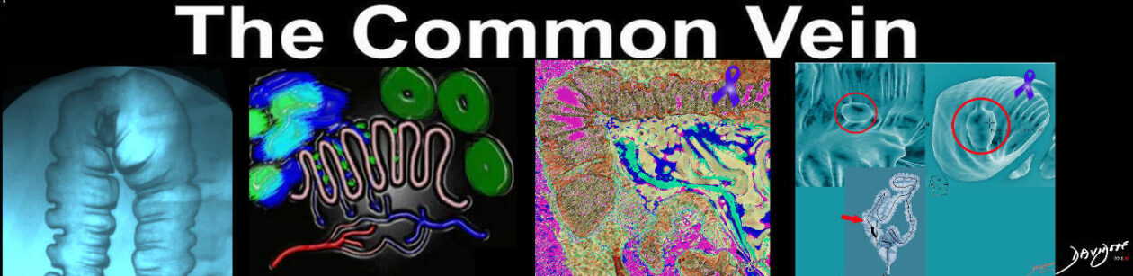
The radiology of acute ulcerative colitis was discussed in the section on diarrhea and is represented below again to correlate with the pathology image above.
Chronic ulcerative colitis shows thickening, shortening rigidity sometimes becoming stiff and straight like a lead pipe. There is also accumulation of fat in the submucosa as well as in the pericolonic region.
|