Lymphatic Drainage
The Common Vein
Copyright 2007
Introduction
The lymph nodes are named mostly according o the vessels along which they travel. Thus the names include the ileocolic, right colic, middle colic, left colic, sigmoid, superior rectal, middle rectal, and inferior rectal lymph nodes. These in turn drain to the appropriate group of nodes situated at the origin of main mesenteric vessels including the SMA, IMA and iliac nodal stations. The nodes that are along the mesenteric border where the marginal artery is located are called the paracolic nodes. In addition there are two special groups of nodes around the cecum. One group is called prececal and the second is called retrocecal – both named according to their position with respect to the cecum.
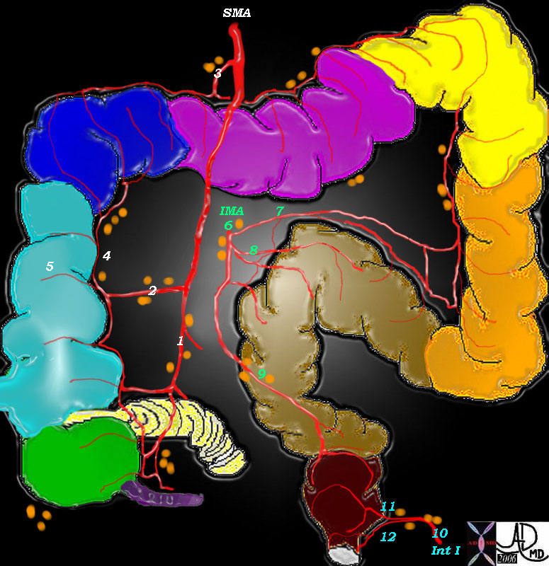 Lymph nodes of the colon Lymph nodes of the colon |
| The lymph nodes are named according to the blood vessels with which they travel. Thus there is the ileocolic group (1) right colic (2), middle colic (3) left colic (7), sigmoid (8), superior rectal (9), middle rectal (11), and inferior rectal (12). These nodes drain into the regional halfway houses for the nodes including the SMA, IMA and iliac groups. The paracolic groups are located along the marginal artery (4) and the prececal and retrocecal are seen in relation to the cecum.
Courtesy Ashley Davidoff MD
12005b017 |
The solitary lymphatic nodules of submucosal lymphoid tissue in the large intestine are most abundant in the cecum and appendix, but are also found throughout the rest of the colon.
Applied Anatomy
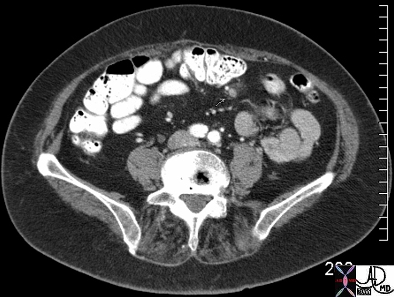 Colon carcinoma with an inflamed node Colon carcinoma with an inflamed node |
| This middle aged female with colon carcinoma has a paracolic lymph node (orange) that is less than 1cms, but it has a halo of induration eccentric at 2o’clock around it. Peritoneal metastases were also found and therefore it is very likely that the small node seen in this image was metastatic given its unusual appearance.
Courtesy Ashley Davidoff MD
45059 45059b01 |
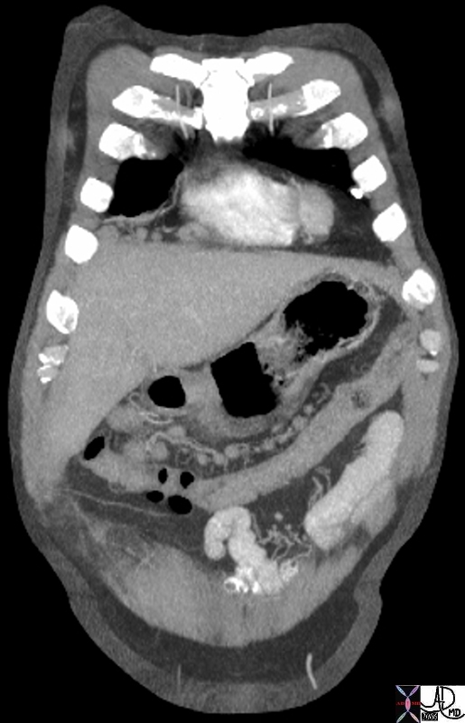 Paracolic lymph nodes Paracolic lymph nodes |
| The paracolic lymph nodes (orange overlay) travel with the marginal artery along the mesenteric border of the colon, and are seen as sub centimeter soft tissue densities along the upper border of the decompressed colon. They are situated in this case within the transverse mesocolon along the middle colic territory. These are not usually as large as this but were prominent in this patient who had bulky lymphomatous nodes along the superior mesenteric vessels.
Courtesy Ashley Davidoff MD
45565 45565b01 |
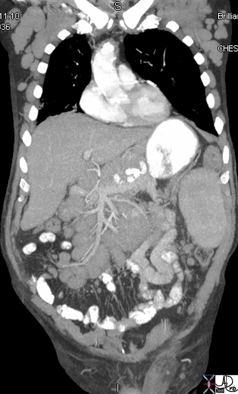 Mesenteric nodes. Mesenteric nodes. |
| This elderly man has lymphoma and the enlarged lymph nodes are clustered around the SMV (maroon) and SMA (red) like a bunch of grapes. Smaller nodes are also seen in the small bowel mesentery and along the left colic and left paracolic region.
Courtesy Ashley Davidoff MD
45568b05 |
|